Bioanalytical Research
Bioanalytical Research Group
Results
The first protein used was the monomeric channel protein ompG. The molecular structure for ompG predicts that a membrane with ompG channels would show 4 nm wide pores. The transmission electron microscopic picture of the membrane preparation shadowed with platinum showed such a layer extended over the entire 3 mm wide preparation area.

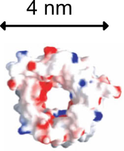
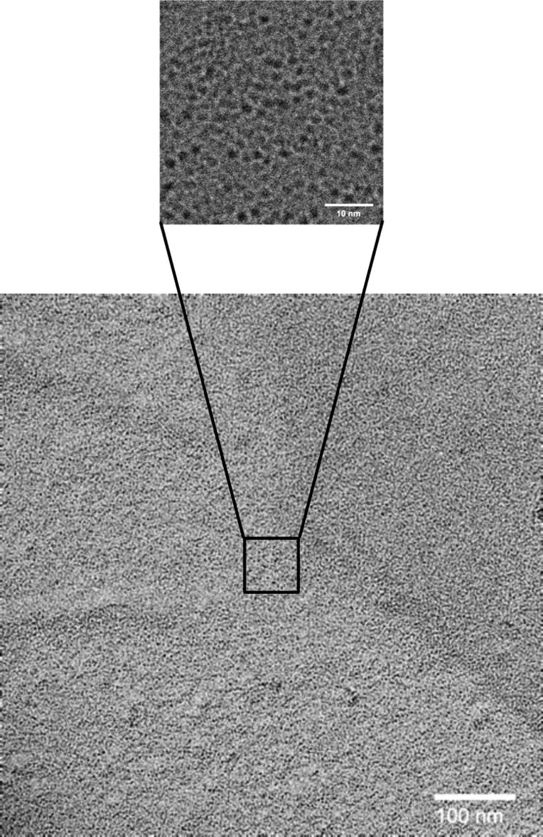
Transmission electron microscopic picture of an extended ompG containing membrane. Platinum shadowing was used to generate contrast.

